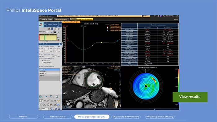
Moduł MR Cardiac do precyzyjnej oceny czynnościowej i ilościowej badań serca
Poznaj ofertę rozwiązań zaawansowanej wizualizacji w kardiologii - MR Cardiac służących do oceny czynności mięśnia sercowego, jego objętości oraz zbliznowacenia.
Zmiana oblicza zaawansowanej wizualizacji w kardiologii
Zyskaj dostęp do narzędzi umożliwiających kompleksową diagnostykę i monitorowanie chorób serca. Modele 3D, mapy i inne narzędzia do oceny ilościowej pozwalają przyspieszyć analizy i usprawnić procedury diagnostyczne. Odkryj korzyści zaawansowanej diagnostyki obrazowej w pracowni interwencyjnej dzięki integracji pakietu Allura/Azurion Interventional Suite z systemem IntelliSpace Portal, który automatycznie pobiera z portalu dane pacjentów, dla których zaplanowano zabiegi.
- Calcium Scoring
-
CT Calcium Scoring
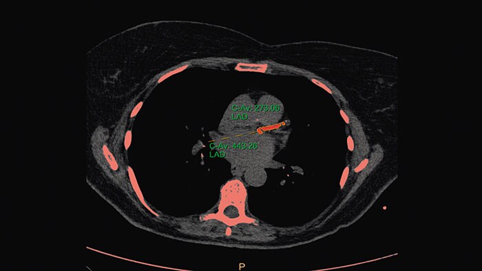
Trójwymiarowa segmentacja zwapnień uruchamiana jednym kliknięciem myszy
Aplikacja umożliwiająca szybką ocenę ilościowej zwapnienia tętnic wieńcowych (CAC) z zastosowaniem obliczania całkowitej masy zwapnień, skali Agatstona oraz obliczania iloczynu liczby wokseli zawierających wapń i objętości jednego woksela. Aplikacja umożliwia zautomatyzowaną dystrybucję wyników w formie papierowej lub elektronicznej w postaci niestandardowych opisów dostosowanych do potrzeb użytkownika.
Korzyści
- Wykonywana jednym kliknięciem segmentacja 3D i analiza ilościowa zwapnienia tętnic wieńcowych (CAC) z uwzględnieniem obliczania całkowitej masy zwapnień, skali Agatstona oraz obliczania iloczynu liczby wokseli zawierających wapń i objętości jednego woksela.
- Opisy dostosowane do potrzeb placówki obejmujące wyniki pomiarów i wskaźnik zwapnienia tętnic wieńcowych.
- Aplikacja charakteryzuje się możliwością edytowania opisów, a także łatwego tworzenia i dodawania do konfiguracji systemu nowych domyślnych szablonów.
- W procedurze analizy uwzględniono ocenę ryzyka na podstawie badania MESA (ang. Multi-Ethnic Study of Atherosclerosis), a jej wynik jest włączany do opisu.
- Cardiac Plaque Assessment
-
CT Cardiac Plaque Assessment
Ocena blaszki miażdżycowej w naczyniach wieńcowych
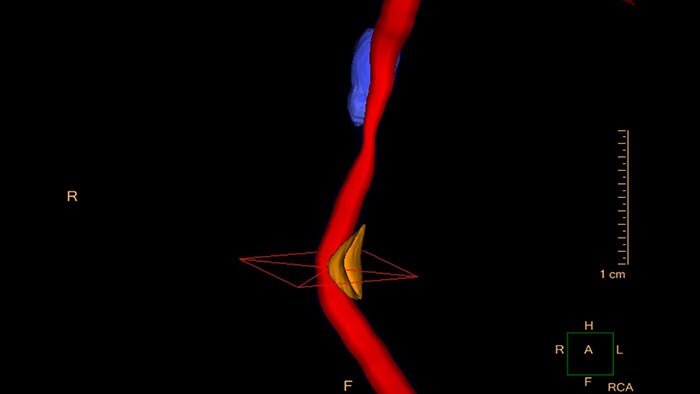
Aplikacja CT Cardiac Plaque Assessment umożliwia ocenę miejsc nagromadzenia blaszki miażdżycowej oraz ocenę ilościową i analizę blaszki miażdżycowej w naczyniach wieńcowych.
Korzyści
- Funkcje oceny ilościowej i charakterystyki blaszki miażdżycowej w naczyniach wieńcowych na podstawie danych wielodetektorowej tomografii komputerowej (MDCT).
- Cardiac Viewer
-
CT Cardiac Viewer
Szybka wizualizacja mięśnia sercowego
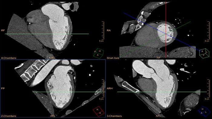
Kompletny zestaw narzędzi do szybkiej wizualizacji jednej lub kilku faz cyklu pracy serca.
Korzyści
- Synchronizacja wielu faz cyklu pracy serca z interaktywnymi narzędziami do prezentacji warstw w widokach MIP w celach poglądowych.
- Funkcja usuwania żeber z obrazów CT klatki piersiowej pozwala na trójwymiarową rekonstrukcję objętości anatomicznej serca oraz połączonych z nim dużych naczyń krwionośnych po anatomicznym usunięciu żeber na potrzeby różnych zadań klinicznych i protokołów skanowania. Funkcja ta pomaga w wizualizacji złożonych struktur anatomicznych oraz w udostępnianiu wyników (np. chirurgom).
- Comprehensive Cardiac Analysis (CCA)
-
CT Comprehensive Cardiac Analysis (CCA)
Kompleksowa analiza badań CT serca
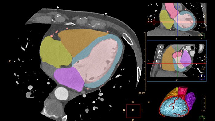
Ta aplikacja pozwala na automatyczną segmentację całego serca w oparciu o model trójwymiarowy, jak również segmentację tętnic wieńcowych bez udziału użytkownika, umożliwiając automatyczną ekstrakcję i wizualizację całego drzewa naczyń wieńcowych.
Korzyści
- Możliwa jest wizualizacja całego drzewa naczyń wieńcowych, analiza morfologiczna światła naczynia oraz analiza średnicy obszaru niezablokowanego światła naczynia.
- Możliwość przeprowadzania analizy czynnościowej komór oraz trójwymiarowej wizualizacji morfologii jam i zastawek serca z wykorzystaniem trybu dynamicznej sekwencji filmowej. Dodatkowo dostępne są obliczenia objętości fali zwrotnej i wskaźnika frakcji, objętości prawej/lewej komory serca we wczesnej i późnej (czynnej i biernej) fazie napełniania oraz współczynnika napełniania lewej komory serca we wczesnej fazie w porównaniu z fazą późną.
- MI Fusion
-
CT-MI Fusion
Fuzja obrazów CT i NM serca
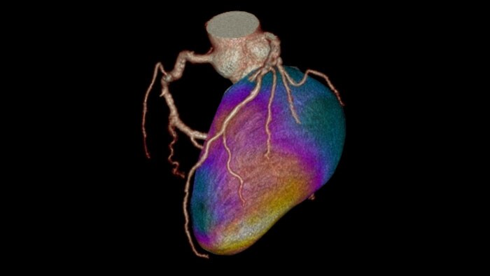
Aplikacja Cardiac CT-NM Fusion oferuje obsługę obrazowania perfuzji mięśnia sercowego (MPI).
Korzyści
- Możliwość wczytywania danych CT wraz ze zbiorami danych NM z obrazowania bramkowanego i niebramkowanego w spoczynku oraz obrazowania bramkowanego i niebramkowanego w obciążeniu.
- Obrazy NM są wyświetlane w projekcjach w osi krótkiej i dwóch płaszczyznach w osi długiej.
- Osie są definiowane na podstawie badania CT.
- Dynamic Myocardial Perfusion (DMP)
-
CT Dynamic Myocardial Perfusion (DMP)
Dynamiczne mapy barwne pozwalają na ocenę ryzyka choroby wieńcowej
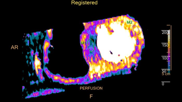
Ta aplikacja służy do wizualizacji, oceny diagnostycznej i oceny ilościowej obrazów mięśnia sercowego ze szczególnym uwzględnieniem lewej komory. Zapewnia w szczególności pomiary ilościowe przepływu krwi przez serce na obrazach CT, w tym identyfikację obszarów mięśnia sercowego o obniżonej perfuzji, mogących wskazywać na niedokrwienie. Aplikacja obsługuje obrazy CT wykonane w projekcji osiowej, bramkowane sygnałem EKG, obejmujące wiele ujęć tego samego obszaru mięśnia sercowego w odstępach czasowych. Aplikacja CT Dynamic Myocardial Perfusion wyświetla wyniki w postaci obrazu złożonego (jeden obraz obliczony z serii obrazów wykonanych w jednym miejscu w określonym czasie).
Korzyści
- Prezentacja przepływu i objętości krwi w obrębie mięśnia sercowego w postaci mapy kodowanej kolorem.
- Analiza parametrów perfuzji na podstawie mapy kodowanej kolorem i krzywych zależności gęstości do czasu.
- Wyrównanie przestrzenne zapewniające dokładniejszą ocenę ilościową perfuzji mięśnia sercowego.
- EP Planning
-
CT EP Planning
Planowanie zabiegów elektrofizjologicznych
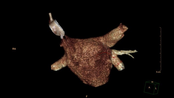
Aplikacja CT EP Planning pozwala lekarzowi na szybką identyfikację struktur anatomicznych istotnych z punktu widzenia zabiegu elektrofizjologicznego.
Korzyści
- Dostęp do całościowej oceny żył płucnych oraz anatomii lewego przedsionka i uszka pozwalający lekarzowi na identyfikację struktur mogących utrudnić wykonanie zabiegu elektrofizjologicznego.
- Myocardial Defect Assessment
-
CT Myocardial Defect Assessment
Ocena zmian w mięśniu sercowym
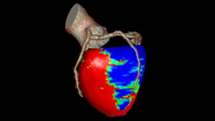
Aplikacja ta pozwala na wizualną i ilościową ocenę posegmentowanych obszarów zmian o niskim współczynniku tłumienia promieniowania w mięśniu sercowym na podstawie pojedynczego, bramkowanego skanu CTA (tomografia spiralna serca z bramkowaniem retrospektywnym lub protokół skanowania Step and Shoot Cardiac).
Aplikację CT Myocardial Defect Assessment opracowano na podstawie zaawansowanej, automatycznej, opartej na modelu techniki segmentacji całego serca pochodzącej z aplikacji CT Comprehensive Cardiac Analysis.
Korzyści
- Możliwości wizualnej oceny obszarów zmian o niskim współczynniku tłumienia promieniowania w obrębie lewej komory serca z wykorzystaniem:
– map kodowanych kolorem w projekcjach w osi krótkiej,
– map segmentacji w projekcjach w osi krótkiej i map biegunowych wyświetlanych razem z obrazami referencyjnymi w projekcjach w osi długiej,
– objętościowej wizualizacji tętnic wieńcowych wraz z mapami segmentacji wyświetlanymi jako nakładki na powierzchnię trójwymiarowego modelu serca.
- Spectral Advanced Vessel Analysis
-
CT Spectral Advanced Vessel Analysis
Funkcja tomografu IQon Spectral CT
Korzyści
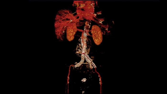
- Usuwanie kości przy różnych poziomach energii.
- Wykresy spektralne umożliwiające charakteryzowanie blaszki miażdżycowej i zwężenia naczyń.
- Porównanie wyników uzyskanych przy różnych poziomach energii.
- Ocena stopnia okluzji światła naczynia.
- Spectral Comprehensive Cardiac Analysis
-
CT Spectral Comprehensive Cardiac Analysis
Funkcja tomografu IQon Spectral CT
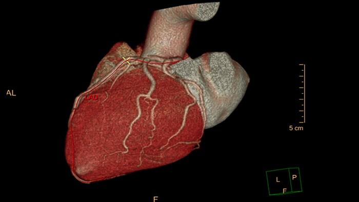
Umożliwia przeprowadzanie segmentacji serca przy różnych poziomach energii, porównywanie krzywych naczyń z różnymi mapami spektralnymi oraz poprawia wizualną ocenę drożności naczynia wieńcowego.
Korzyści
- Automatyczna segmentacja jam serca i naczyń wieńcowych z użyciem obrazów monoenergetycznych.
- Redukcja artefaktów utwardzania wiązki na potrzeby wizualizacji deficytów w perfuzji mięśnia sercowego oraz blaszki miażdżycowej.
- Spectral Light Magic Glass
-
Funkcja Light Magic Glass w tomografii spektralnej
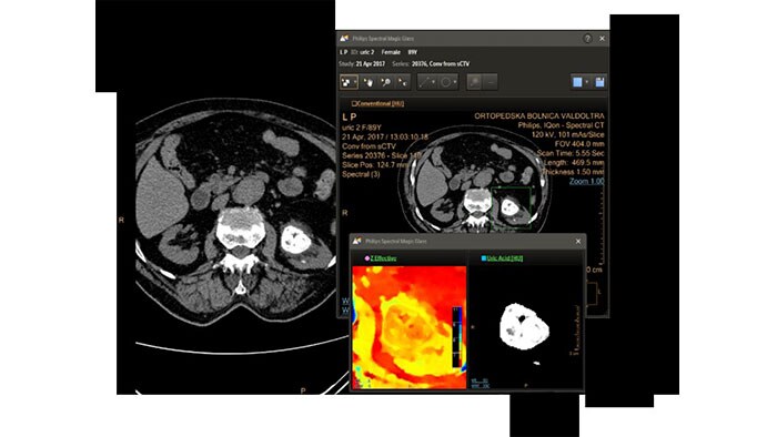
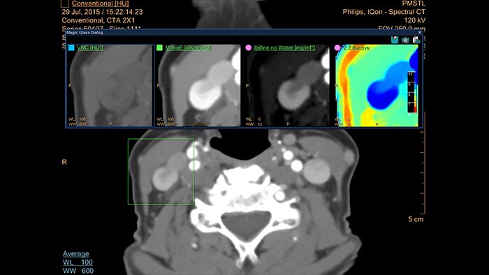
Umożliwia retrospektywne wykorzystanie danych spektralnych zapisanych jako obrazy SBI. Możliwe jest przeglądanie danych spektralnych oraz identyfikacja najbardziej istotnych wyników do załadowania do konwencjonalnej aplikacji CT w celu przeprowadzenia rutynowej analizy — nawet do aplikacji, które nie zostały opracowane pod kątem obsługi danych spektralnych.
Korzyści
- Możliwe jest przeglądanie danych spektralnych oraz identyfikacja najbardziej istotnych wyników do załadowania do konwencjonalnej aplikacji CT w celu przeprowadzenia rutynowej analizy — nawet do aplikacji, które nie zostały opracowane pod kątem obsługi danych spektralnych:
– Aplikacja do wirtualnej kolonoskopii Virtual Colonoscopy
– Aplikacja do analizy obrazów wątroby Liver Analysis
– Przeglądarka obrazów pacjentów po urazach Trauma Viewer (Acute Multifunctional Review)
– Aplikacja do planowania zabiegów TAVI
– Aplikacja do analizy tętnic płuc PAA
– Aplikacja do analizy perfuzji mózgu Brain Perfusion
– Aplikacja do analizy perfuzji narządowej Functional CT (FCT)
- Spectral Magic Glass on PACS
-
Spectral Magic Glass w systemie PACS*
Funkcja tomografu IQon Spectral CT
IQon Spectral CT jest jedynym tomografem wyposażonym w aplikacje CT Spectral Light Magic Glass oraz CT Spectral Magic Glass on PACS, które ułatwiają radiologom przeglądanie i analizowanie wielu warstw danych spektralnych jednocześnie, także z poziomu systemu PACS.
Korzyści
- Możliwość przeprowadzania na żądanie jednoczesnej analizy wielu wyników spektralnych dla obszaru zainteresowania (ROI).
- Integracja z systemem PACS szpitala dostępna dla systemów PACS określonych producentów.
- Możliwość przeglądania wyników analizy spektralnej podczas rutynowego opisywania obrazów.
- Możliwość przeglądania i analizy danych spektralnych w praktycznie każdej lokalizacji w obrębie całej placówki.
*Dostępna w standardzie z opcją CT Spectral w systemie IntelliSpace Portal.
- TAVI Planning
-
CT TAVI Planning
Obrazowanie CT na potrzeby planowania TAVI w zaawansowanej diagnostyce pacjenta
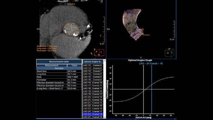
Dostęp do funkcji półautomatycznych pomiarów aorty i zastawki aortalnej przydatnych na etapie planowania zabiegu TAVI.
Korzyści
- Oparta na modelu segmentacja zastawki aortalnej z zastosowaniem automatycznej segmentacji zwapnień i ulepszonej funkcji wykrywania punktów charakterystycznych.
- Pomiary struktur anatomicznych na potrzeby dobrania wielkości narzędzia do zabiegu TAVI.
- Wskazanie właściwego kąta początkowego ramienia C przy umiejscawianiu narzędzia w pracowni hemodynamiki lub na hybrydowej sali operacyjnej.
- Aplikacja przedstawia drogę dostępu naczyniowego, dzięki czemu potencjalnie skraca czas procedur.
- 3D Modeling
-
3D Modeling*
Uproszczony proces tworzenia modeli, zoptymalizowany pod kątem wydruku na drukarkach 3D
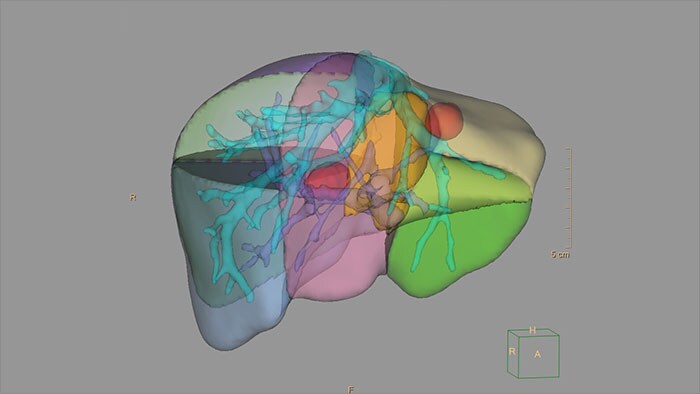
System IntelliSpace Portal zawiera dedykowaną aplikację 3D Modeling przeznaczoną do tworzenia i eksportowania trójwymiarowych modeli. To zintegrowane środowisko programowe zapewnia dostęp do narzędzi segmentacji systemu IntelliSpace Portal zebranych w jednym miejscu w celu uproszczenia przebiegu pracy.
Korzyści
- Obsługa importu wyników segmentacji 3D z innych aplikacji oraz tworzenia własnych niestandardowych modeli bezpośrednio z obrazów DICOM.
- Dostęp do szeregu dostosowanych do potrzeb lekarzy narzędzi rekonstrukcji i edycji, które pomagają zoptymalizować modele pod kątem wydruku 3D, tak aby faktycznie odzwierciedlały one narząd pacjenta.
- Lekarz może wyświetlić podgląd siatki w zestawieniu z oryginalnymi obrazami DICOM i wykonać w czasie rzeczywistym poprawki:
– Aplikacja 3D Modeling umożliwia łatwy eksport plików zestawów danych w standardowych formatach (takich jak STL) oraz w formacie 3D PDF, który można wykorzystać do wymiany informacji w obrębie placówki.
– Dostępność różnych opcji eksportu usprawnia przesyłanie plików do usługi drukowania, jak również w obrębie szpitala na użytek wewnętrzny.
*W Stanach Zjednoczonych modele 3D nie zostały dopuszczone do stosowania w diagnostyce.
- Advanced Vessel Analysis (AVA)
-
Multi Modality Advanced Vessel Analysis (AVA)
Wszechstronna analiza naczyń na potrzeby planowania zabiegów
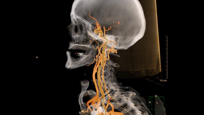
Aplikacja Multi Modality Advanced Vessel Analysis (AVA)* służy do oceny i analizy ilościowej różnych rodzajów zmian naczyniowych uwidocznionych w badaniach CRA i MRA. Narzędzie AVA umożliwia wykonywanie analizy różnymi metodami i oznaczanie różnych zmian naczyniowych, a także ułatwia nawigację w obrębie wielu rozpoznanych nieprawidłowości.
Aplikacja CT Advanced Vessel Analysis (AVA) Stent Planning zapewnia użytkownikowi dostęp do danych ilościowych oraz parametrów stentu w celu wygenerowania informacji niezbędnych do zaplanowania zabiegów interwencyjnych, takich jak EVAR.
Korzyści
- Zaawansowany algorytm usuwania kości umożliwia trójwymiarową wizualizację naczyń.
- Zautomatyzowane narzędzia, takie jak linie środkowe, oznaczanie naczyń oraz określanie wewnętrznychi zewnętrznych konturów światła naczynia, a także funkcja automatycznego tworzenia serii Automatic Series Creation (ASC), pozwalają na szybsze uzyskanie wyników końcowych i poprawiają ich spójność.
- Łatwe przechodzenie między wieloma wynikami i możliwość eksportowania dostosowanych do potrzeb, szczegółowych opisów do systemu RIS lub PACS po zakończeniu pracy z badaniem.
- Definiowane przez użytkownika opcje zaawansowanej analizy naczyń na potrzeby planowania zabiegów.
- Narzędzia edycji tkanek dostępne z poziomu pływającego paska narzędzi otwartego w wybranym polu obrazu.
*W porównaniu ze stacją diagnostyczną EBW w wersji 4.x firmy Philips.
- Cardiac
-
MR Cardiac
Kompleksowy przegląd i analiza ilościowa badań MR serca.
Aplikacja MR Cardiac umożliwia wizualizację jednej, kilku lub wszystkich serii obrazów serca z użyciem protokołów wyświetlania, w tym z synchronizacją z fazami cyklu pracy serca. Intuicyjna dedykowania przeglądarka umożliwia szybkie zapoznanie się z ogólnymi wynikami badań serca oraz zadecydowanie, która analiza jest wymagana w pierwszej kolejności. Możliwe jest dokonanie wizualnej oceny przy użyciu wykresu kołowego (ang. bull's eye) zgodnego z zaleceniami AHA.
Pakiet pozwala na wykonywanie kompleksowej objętościowej analizy czynności komór uwzględniającej parametry takie jak frakcja wyrzutowa, ruch ścian serca, ich grubość i pogrubienie. Oferuje ona identyfikację wzmocnienia przestrzennego w oparciu o zmiany natężenia sygnału oraz funkcję zakładki, która pozwala oznaczyć ramką dowolny widok danych, które są istotne i wymagają zapisania lub przekazania innym lekarzom. Aplikacja MR Cardiac umożliwia również przeprowadzanie szybkiej analizy czynnościowej metodą ALEF (Areal Length Ejection Fraction).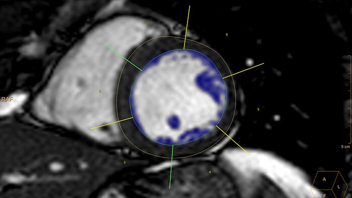
Korzyści
- Etykietowanie serii obrazów MR serca umożliwia dokonywanie ich szybkich przeglądów.
- Łatwiejsze przeglądanie obrazów dzięki łączeniu faz cyklu pracy serca i układów przestrzennych.
- Narzędzia do półautomatycznej i ręcznej segmentacji lewej i prawej komory do analizy badań funkcjonalnych serca.
- Możliwość wyświetlenia wszystkich warstw serii w krótkiej osi serca w jednym widoku.
- Możliwość edytowania danych źródłowych i ponownego obliczania map parametrycznych przez dopasowanie krzywej metodą najmniejszych kwadratów.
- Cardiac Quantitative Mapping
-
MR Cardiac Quantitative Mapping
Ocena cech tkanki mięśnia sercowego
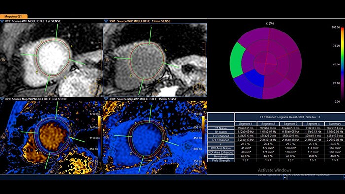
Aplikacja MR Cardiac Quantitative Mapping ułatwia ocenę i przegląd cech tkanki mięśnia sercowego w wielu definiowanych przez użytkownika, uwzględniających natężenie pola, tabelach przeglądowych.
Korzyści
- Globalne i rozproszone zmiany patologiczne mięśnia sercowego można przeglądać na mapach T1, T2 i T2*.
- Narzędzia do ręcznej i automatycznej korekcji ruchu, potencjalnie zwiększające dokładność obliczeń map.
- Obsługa wielu rodzajów akwizycji: Molli, shMolli, SASHA, T2prep.
- Cardiac Temporal Enhancement
-
MR Cardiac Temporal Enhancement
Identyfikacja zmian natężenia sygnału w dynamicznych badaniach MR serca
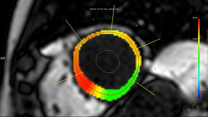
Aplikacja MR Cardiac Temporal Enhancement ułatwia analizę przepływu krwi przez serce dzięki wykorzystaniu dynamicznych obrazów serca (wielowarstwowych, wielodynamicznych). Aplikacja generuje wyniki w różnych formatach wyjściowych, umożliwiając przegląd zidentyfikowanych zmian czasowych.
Korzyści
- Opcje ręcznej segmentacji i edycji lewej komory; opcjonalna zautomatyzowana korekcja afiniczna lub sztywna w celu kompensacji ruchu oddechowego.
- Zintegrowana funkcja porównania danych z badań spoczynkowych i obciążeniowych.
- Różne formaty danych wyjściowych, w tym wykresy natężenia w czasie, wykresy tarczowe, wykresy wzmocnienia w czasie oraz kodowane kolorem nakładki na obrazy anatomiczne.
- Cardiac Whole Heart
-
MR Cardiac Whole Heart
Szczegółowa wizualizacja 3D posegmentowanego serca
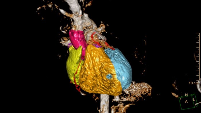
Aplikacja MR Cardiac Whole Heart przeprowadza automatyczną segmentację serca na poszczególne struktury składowe (takie jak lewa komora, prawa komora, przedsionki i naczynia wieńcowe).
Korzyści
- Wyniki mogą być prezentowane w postaci wysokiej jakości wizualizacji 3D, również w formatach eksportu danych 3D (np. STL) na potrzeby wydruku modeli 3D.
- Przebieg pracy polegający na tworzeniu nowego segmentu tkankowego obsługuje segmentację opartą na maskowaniu i obszarach rozrostowych.
- Segmentacja oparta na obszarach rozrostowych.
- Minimalny udział użytkownika w procesie dzięki udoskonalonej segmentacji 3D.
- Tworzenie jednego widoku/modelu 3D na podstawie danych pozyskanych w różnych seriach i przy różnych parametrach dynamicznych — wsparcie przy podejmowaniu decyzji dotyczących leczenia złożonych struktur hemodynamicznych.
- Przygotowanie i eksportowanie modeli 3D ze zdefiniowanym przez użytkownika wygładzeniem i stopniem nieprzezroczystości, w formacie odpowiednim do wydruku 3D i obsługiwanym przez oprogramowania do nawigacji chirurgicznej.
- QFlow
-
MR QFlow
Wizualizacja i analiza ilościowa dynamiki przepływu krwi
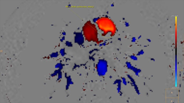
Aplikacja MR QFlow umożliwia wizualizację i ocenę ilościową danych przepływu. Tworzone są dwuwymiarowe, kodowane kolorem mapy przepływu w formie nakładek na obrazy anatomiczne, wykorzystywane na przykład do obliczania objętości wyrzutowych.
Aplikacja QFlow jest również integralną częścią pakietu MR Cardiac Suite, umożliwiając opisywanie badań w połączeniu z innymi narzędziami analitycznymi, takimi jak narzędzie do oceny czynnościowej.
Korzyści
- Funkcja automatycznego wykrywania konturów dużych naczyń na potrzeby analizy przepływu w tych strukturach.
- Korekcja tła umożliwia wykonanie korekcji przesunięcia wymaganej w przypadku danych oceny ilościowej przepływu uzyskanych przy użyciu systemów MR niektórych producentów.
- Możliwość zintegrowania aplikacji QFlow w pakiecie MR Cardiac Suite.
- Możliwość porównywania wyników analizy przepływu z czynnością serca w ramach JEDNEGO pakietu aplikacji.
- Wspólne opisywanie wyników analizy QFlow oraz analizy czynnościowej (i innych analiz).
- Astonish Reconstruction
-
NM Astonish Reconstruction
NM Astonish Reconstruction to zaawansowany algorytm rekonstrukcji, w którym wykorzystano opracowaną przez firmę Philips technikę zharmonizowanego, podwójnego filtrowania, mającą na celu ograniczenie szumów oraz poprawę rozdzielczości i jednorodności rekonstruowanych obrazów. Ponadto w celu korekcji tłumienia w połączeniu z aplikacją NM Astonish Reconstruction można stosować mapę tłumienia wygenerowaną z użyciem tomografii komputerowej. Dzięki poprawie stosunku sygnału do szumu można uzyskać obrazy odpowiedniej jakości przy potencjalnym skróceniu czasu skanowania SPECT, co pozwala zwiększyć liczbę wykonywanych badań, poprawić komfort pacjentów podczas badania i ograniczyć powstawanie artefaktów ruchowych.
Korzyści
- Liczne zalety kliniczne, w tym wyższa rozdzielczość obrazów i lepsza wydajność pracy.
- Możliwość obrazowania SPECT serca w czasie skróconym o połowę z użyciem radionuklidów TI-201, In-111, Ga-67, I-123 lub I-131 za pomocą obsługiwanych systemów firmy Philips w celu zwiększenia wydajności pracy przy zachowaniu wysokiej jakości obrazów.
- Narzędzie wspomagające pewność i dokładność diagnostyczną.
- Możliwość stosowania w badaniach SPECT wykorzystujących radioznacznik Tc-99m, najczęściej stosowany znacznik w procedurach obrazowania molekularnego.
- Wyłączna zgodność z następującymi kamerami firmy Philips: CardioMD (oprogramowanie do akwizycji w wer. 2.x), Forte, BrightView, BrightView X, BrightView XCT, SkyLight i Precedence.
- Cedars-Sinai Cardiac Suite 2017
-
NM Cedars Sinai Cardiac Suite 2015*
Zaawansowana ocena mięśnia sercowego
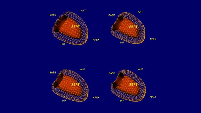
Pakiet NM Cedars Sinai Cardiac Suite 2015 oferuje kompleksowe narzędzia do oceny ilościowej mięśnia sercowego przeznaczone do przetwarzania danych z bramkowanych badań SPECT, badań perfuzji i scyntygrafii puli krwi wykonywanych techniką SPECT oraz ilościowych badań PET. Szeroko znany lekarzom na całym świecie pakiet aplikacji Cedars-Sinai Cardiac Suite 2015 umożliwia sprawne opisywanie badań z dostępną wyłącznie w tym rozwiązaniu oceną perfuzji i czynności.
Korzyści
- Ocena ilościowa prawej komory: zautomatyzowane konturowanie prawej komory, ocena ilościowa i analiza.
- Edytor defektów mapy biegunowej perfuzji: umożliwia użytkownikom ręczne edytowanie map biegunowych.
- Funkcja DataView: dostosowywane przez użytkownika układy wyświetlania.
- Udoskonalony algorytm analizy fazowej, funkcja Smart Launch oraz edytor palety kolorów.
*Produkt niedostępny w sprzedaży w niektórych krajach. Należy sprawdzić dostępność w danym kraju.
- Corridor4DM 2018
-
NM Corridor4DM 2016*
Ocena, przegląd i tworzenie raportów z badań SPECT i PET serca i naczyń
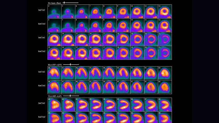
Aplikacja NM Corridor4DM 2016* jest przeznaczona do zaawansowanej oceny ilościowej układu sercowo-naczyniowego oraz wyświetlania obrazów. Oferuje inteligentną organizację pracy oraz opcje kontroli jakości. Korzystając z wielu ekranów przeglądu ze zintegrowanymi narzędziami tworzenia opisów oraz niestandardowymi szablonami, można przeprowadzać ocenę ilościową perfuzji mięśnia sercowego oraz jego czynności i żywotności. Aplikacja NM Corridor4DM w wersji 2016 zawiera ponadto narzędzia do obliczania i oceny ilościowej powierzchni lewej komory, dodatkowe bazy danych referencyjnych oraz rekonstrukcję GEMS Evolution SPECT.
Korzyści
- Narzędzia do analizy ilościowej, wyświetlania i opisywania perfuzji i czynności mięśnia sercowego na podstawie badań SPECT i PET, metabolizmu FDG w badaniach PET oraz badań scyntygraficznych puli krwi techniką SPECT w pojedynczej, konfigurowalnej aplikacji.
- Narzędzia do pozyskiwania i przeglądania statycznych oraz wieloklatkowych obrazów SC w formacie DICOM.
- Łatwo konfigurowalne ustawienia różnych procedur roboczych, protokołów i preferencji.
- Ocena ilościowa rezerwy przepływu wieńcowego (CFR) dla chlorku rubidu (Rb-82) i amoniaku (N-13).
- Narzędzia do obliczania i oceny ilościowej powierzchni lewej komory.
- Udoskonalone narzędzia wyświetlania.
- Dodatkowe bazy danych wartości prawidłowych do obsługi rekonstrukcji GEMS Evolution SPECT.
- Zaktualizowane narzędzia do pozyskiwania i przeglądania statycznych oraz wieloklatkowych obrazów SC w formacie DICOM.
* Corridor4DM jest zastrzeżonym znakiem towarowym firmy Invia, LLC.
- Emory Cardiac Toolbox (ECTb) SyncTool
-
Emory Cardiac Toolbox (ECTb) SyncTool*
Ocena dyssynchronii mechanicznej serca
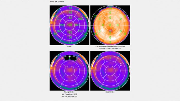
Aplikacja Emory Cardiac Toolbox (ECTb) SyncTool umożliwia obiektywną ocenę dyssynchronii lewokomorowej na podstawie analizy faz cyklu pracy serca.
Korzyści
- Dostęp do dodatkowych informacji prognostycznych, które można uzyskać z trójwymiarowych obrazów perfuzji, dotyczących przykładowo obecności tkanki bliznowatej i jej umiejscowienia.
- Ekran przeglądu danych obejmujący prezentację map biegunowych faz cyklu pracy serca, histogramy faz oraz podsumowanie analizy pogrubienia skurczowego ściany z uwzględnieniem fazy szczytowej i odchylenia standardowego rozkładu faz.
*Emory Cardiac Toolbox, ECTb, HeartFusion i SyncTool są zarejestrowanymi znakami towarowymi Emory University.
- Emory Cardiac Toolbox (ECTb) HeartFusion
-
Emory Cardiac Toolbox (ECTb) HeartFusion*
Ocena połączonych obrazów budowy drzewa wieńcowego
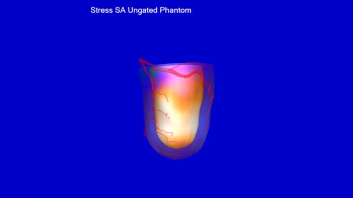
Aplikacja Emory Cardiac Toolbox (ECTb) HeartFusion umożliwia łączenie obrazów drzewa naczyń wieńcowych pacjenta otrzymanych na podstawie angiogramów CT serca z obrazami NM perfuzji.
Korzyści
- Narzędzie umożliwiające skorelowanie stenozy z upośledzeniem perfuzji i identyfikację zagrożonej masy mięśniowej.
*Emory Cardiac Toolbox, ECTb, HeartFusion i SyncTool są zarejestrowanymi znakami towarowymi Emory University.
- Emory Cardiac Toolbox (ECTb) v4.2
-
Emory Cardiac Toolbox (ECTb) w wer. 4.1*
Analiza kardiologiczna
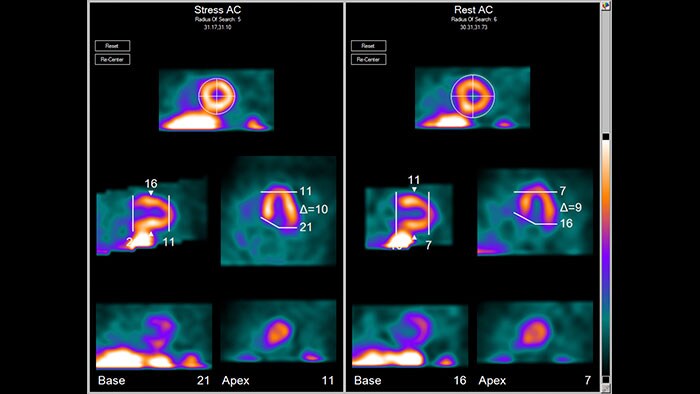
Aplikacja NM Emory Cardiac Toolbox (ECTb) w wer 4.1 oferuje zaawansowane narzędzia do analizy SPECT i PET mięśnia sercowego, w tym porównanie danych perfuzyjnych z danymi dotyczącymi żywotności, możliwość prezentacji obrazów trójwymiarowych z nakładkami naczyń wieńcowych oraz bramkowaną trójwymiarową sekwencją filmową, granice normy dla dopasowania/niedopasowania środka kontrastowego, a także opcjonalną analizę fazową dla ruchu ścian serca i ocenę pogrubienia.
Korzyści
- Nowa opcja SmartReport — zautomatyzowane raportowanie strukturalne dedykowane dla kardiologii nuklearnej.
- Reorientacja transosiowa.
- Ogólna poprawa wydajności.
- Rozszerzona analiza dyssynchronii skurczowej.
- Analiza dyssynchronii rozkurczowej.
*Emory Cardiac Toolbox, ECTb, HeartFusion i SyncTool są zarejestrowanymi znakami towarowymi Emory University.
- Mirada Viewer
-
NM Mirada Viewer
Większy komfort opisywania badań NM
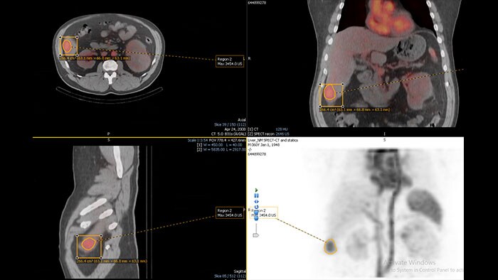
Rozwiązanie zaprojektowane z myślą o sprostaniu wyzwaniom klinicznym i zwiększeniu wydajności podczas przeglądu obrazów PET-CT, SPECT, SPECT-CT i obrazów planarnych. Zoptymalizowany przebieg pracy pozwala na obsługę wielu badań i analizę ilościową zaobserwowanych zmian.
Korzyści
- Szybkie i konfigurowalne protokoły umożliwiające efektywne opisywanie badań.
- Monitorowanie zmian chorobowych i odpowiedzi na leczenie.
- Eksportowanie danych w postaci tabelarycznej i graficznej.
- Rejestracja obrazów PET-CT i PET-CT-MR.
- Rozwiązanie obsługujące informacje pochodzące z systemów różnych producentów*.
*Szczegółowe informacje na temat obsługiwanych rozwiązań poszczególnych producentów można uzyskać, kontaktując się z lokalnym przedstawicielem firmy Philips.
- NM Review
-
NM Review
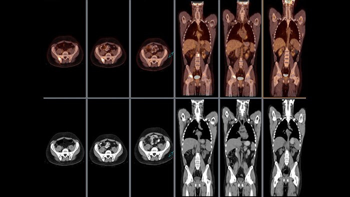
Aplikacja NM Review zapewnia zaawansowane środowisko przeglądania i analizy obrazów NM oraz wykonanych innymi metodami obrazowania na potrzeby klinicznej oceny planarnych badań NM, badań SPECT, SPECT/CT, PET/CT oraz PET/MR.
Korzyści
- Wybór układów pozwala użytkownikowi definiować różne układy dla różnych ustawień predefiniowanych. Dla każdego ustawienia predefiniowanego użytkownik ma do dyspozycji 4 różne układy
- Wyświetlanie widoków MPR, MIP i połączonych trójwymiarowych obrazów objętości.
- Opcja przewijania w trybie ciągłym.
- Pomiary dwu- i trójwymiarowe na podstawie standaryzowanej wartości wychwytu (SUV): SUV Body Weight (masa ciała), SUV Lean Body Mass (beztłuszczowa masa ciała), SUV Body Surface Area (pole powierzchni ciała) i SUV Body Mass Index (wskaźnik masy ciała).
- Zautomatyzowana trójwymiarowa segmentacja zmian w oparciu o wartość SUV lub odsetek maksymalnej wartości SUV oraz funkcja eksportu trójwymiarowych obrysów w formacie DICOM-RT Structure
- Ustawienia formatu w systemach planowania radioterapii.
- US Q-App General Imaging 3D Quantification
-
US Q-App General Imaging 3D Quantification (GI3DQ)
Zaawansowana wizualizacja i ocena lub pomiar objętości na obrazach ultrasonograficznych
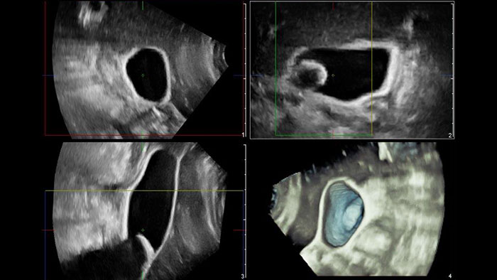
Aplikacja US Q-App General Imaging 3D Quantification (GI3DQ) zapewnia zaawansowane narzędzia do przeglądania, przetwarzania i oceny ilościowej zestawów danych 3D. Dostępne są zaawansowane funkcje obejmujące rekonstrukcję wielopłaszczyznową, obrazowanie tomograficzne iSlice i rekonstrukcje objętościowe, a także pomiary wolumetryczne z zastosowaniem wielu różnych metod, uwzględniających także użycie narzędzi półautomatycznych. Wyniki uzyskane w taki sposób można dołączyć do dokumentacji pacjenta.
Korzyści
- Zaawansowane narzędzia do przeglądania, przetwarzania i analizy trójwymiarowych danych ultrasonograficznych.
- Funkcje rekonstrukcji wielopłaszczyznowej, obrazowania tomograficznego iSlice oraz zaawansowanej rekonstrukcji danych.
- Możliwość łatwego wykonywania pomiarów na obrazach 2D i obrazach objętościowych dzięki użyciu półautomatycznych narzędzi analitycznych.
- Zgodność z zestawami danych 3D pozyskanymi za pomocą ultrasonografów EPIQ, Affiniti, iU22, HD15, HD11 i HD9 firmy Philips.
- Viewing (in MMV)
-
Przeglądanie obrazów angiograficznych (w przeglądarce MMV)
Kompleksowe narzędzie do przeglądania danych pozyskanych różnymi metodami obrazowania — w jednej przeglądarce
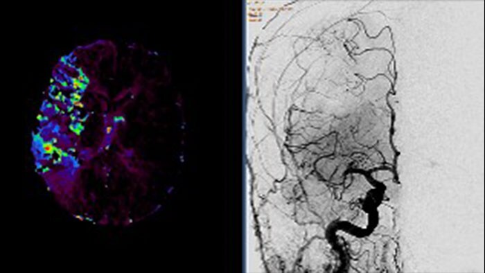
Przeglądarka Multi Modality Viewer (MMV) obsługuje wyświetlanie i post-processing obrazów i serii pozyskanych w badaniach angiograficznych. Dzięki temu można przejrzeć i przeprowadzić analizę obrazów angiograficznych oraz obrazów wykonanych innymi metodami obrazowania w celu uzyskania pełnych informacji na temat stanu pacjenta. Dostępne są następujące funkcje przetwarzania obrazów naczyń (cyfrowa angiografia subtrakcyjna) — subtrakcja, przesuwanie pikseli i rozjaśnianie obrazów w tle. Obrazy kluczowe są umieszczane w ogólnych opisach przeglądarki MMV. Przed zabiegiem interwencyjnym odpowiednie obrazy diagnostyczne (MR i/lub CT) można oznaczyć zakładkami, dzięki czemu zostaną one automatycznie pobrane po wybraniu pacjenta w systemie Allura lub Azurion.
Korzyści
- Dostęp do kompletnych informacji na temat stanu pacjenta z uwzględnieniem wyników badań wykonanych wszystkimi metodami obrazowania — CT/MR/NM/USG/Angio — w jednym środowisku.
- Zaawansowane funkcje wyświetlania i post-processingu (cyfrowa angiografia subtrakcyjna) obrazów i serii pozyskanych w badaniach angiograficznych.
- Funkcje dodawania adnotacji i wykonywania podstawowych pomiarów na obrazach (obraz musi być wstępnie skalibrowany).
- Funkcja iBookmark — automatyczne pobieranie odpowiednich obrazów diagnostycznych (CT i/lub MR) pomocnych podczas zabiegu interwencyjnego.
- Obsługa funkcji opisywania — obrazy kluczowe mogą być wysyłane do ogólnego opisu przeglądarki MMV.
